Beside Vivekanand College, Samarth Nagar, Aurangabad Mon-Sat: 9:30 AM - 5:30 PM

Fundus Photography in Aurangabad

High-resolution digital photography of the retina, optic disc and blood vessels — a permanent record to track your eye health over time.

Understanding Fundus Photography

A Permanent Record of Your Retina

Fundus photography captures a detailed, high-resolution colour image of the back of your eye — the retina, optic disc, macula and blood vessels — giving your doctor a clear, permanent record of your retinal health.

This is especially valuable for tracking changes over time. A single exam gives a snapshot, but comparing fundus photographs taken months or years apart lets our doctors precisely identify new bleeding, growths, or the progression of conditions like diabetic retinopathy or macular degeneration.

At Hazari Eye Hospital, digital fundus photography is integrated with our Cirrus OCT system, giving a complete picture of retinal health in a single visit.
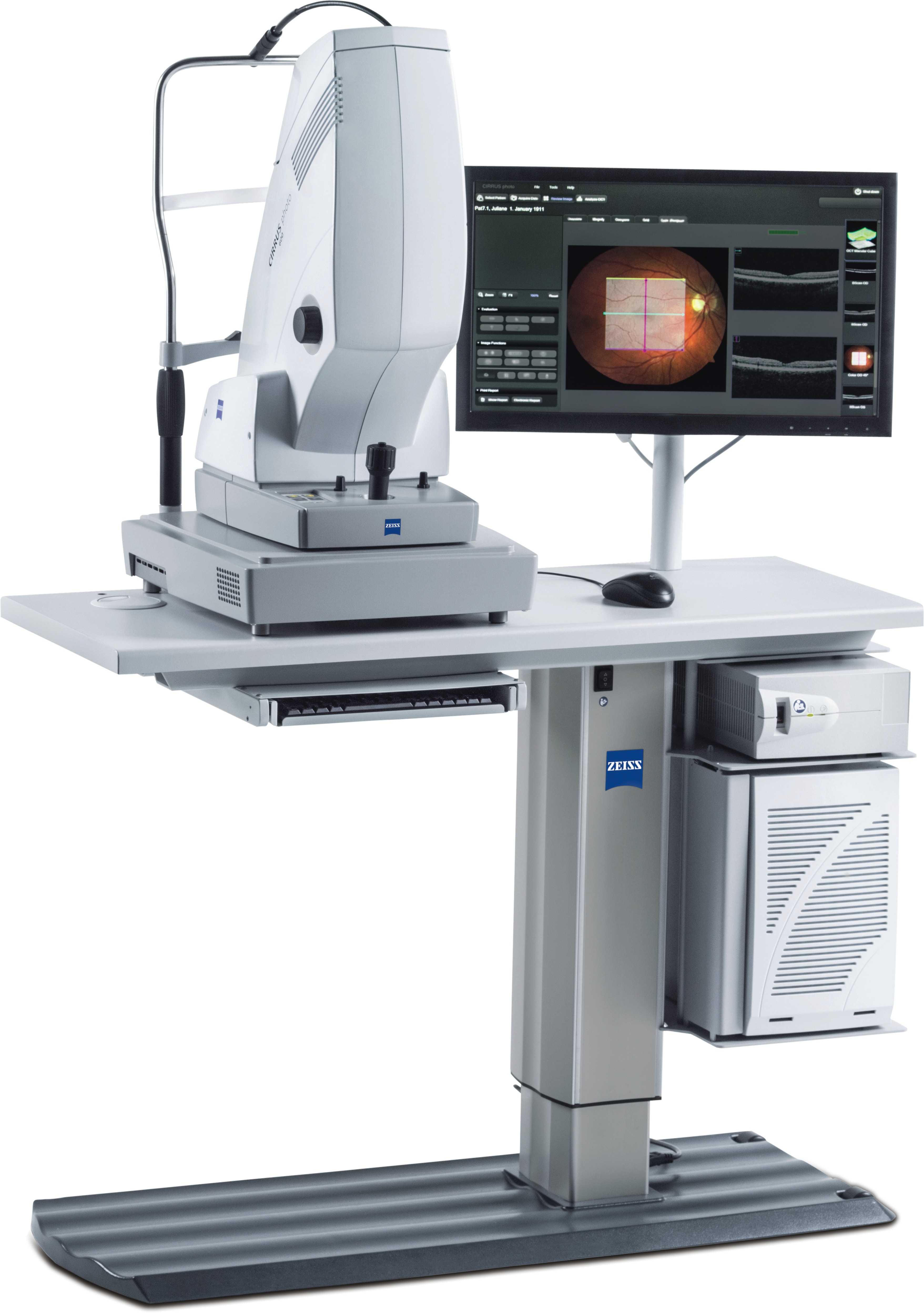
Digital fundus photography at Hazari Eye Hospital
Why It Matters

What Fundus Photography Helps Us Track

Diabetic Retinopathy

Documents bleeding and blood vessel changes for annual comparison.

Optic Disc Health

Tracks the optic nerve appearance relevant to glaucoma monitoring.

Macular Changes

Records early signs of macular degeneration for comparison over time.

Objective Record-Keeping

Gives a factual, comparable record rather than relying on memory alone.

Common Questions

Fundus Photography — FAQs

It's a specialised photograph of the back of your eye (the fundus) — including the retina, optic disc and blood vessels — used to document and monitor your eye health over time.
No. Fundus photography captures a 2D colour photograph of the retinal surface, while OCT captures cross-sectional images showing tissue layers beneath the surface. The two are often used together for a complete picture.
Dilation gives a wider, more detailed view and is often recommended, though some fundus cameras can capture useful images without dilation depending on your examination needs.
Comparing photographs taken months or years apart allows your doctor to precisely track any changes — new bleeding, growths, or progression of a condition — that might not be obvious from memory alone.
No. It's quick, painless and non-contact — you simply position your eye in front of the camera and a bright flash captures the image.
Ready to See Clearly?

Book Your Consultation at Hazari Eye Hospital

Want a documented baseline of your retinal health? Book a fundus photography session today.

Explore More

Other Diagnostic Tests

WhatsApp Call Now